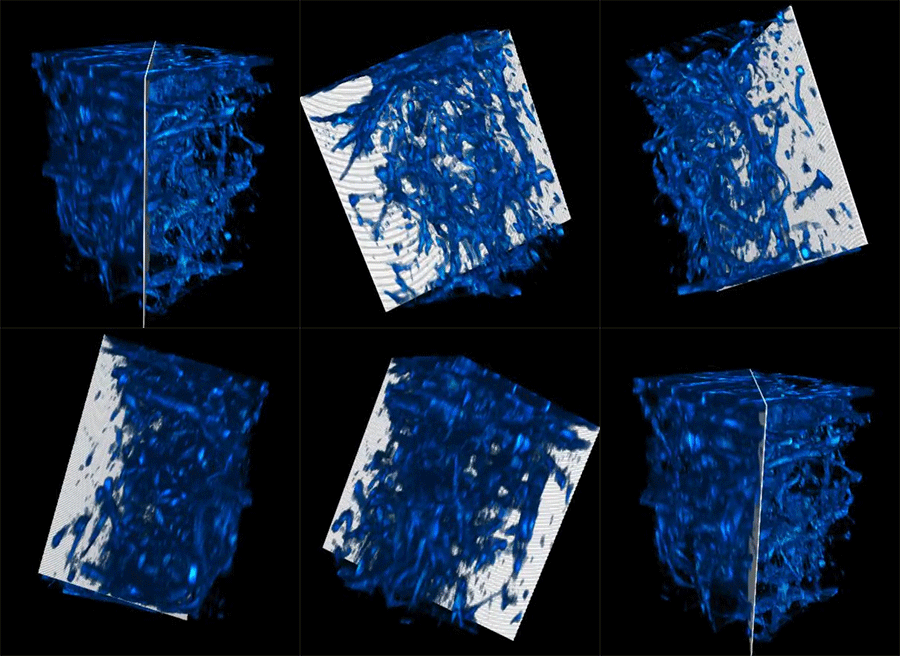
Metabolic imaging is a noninvasive means that allows clinicians and scientists to check residing cells the use of laser mild, which will lend a hand them assess illness development and remedy responses.However mild scatters when it shines into organic tissue, proscribing how deep it could actually penetrate and hampering the decision of captured photographs.Now, MIT researchers have advanced a brand new methodology that greater than doubles the standard intensity restrict of metabolic imaging. Their means additionally boosts imaging speeds, yielding richer and extra detailed photographs.This new methodology does no longer require tissue to be preprocessed, equivalent to through chopping it or staining it with dyes. As an alternative, a specialised laser illuminates deep into the tissue, inflicting sure intrinsic molecules throughout the cells and tissues to emit mild. This gets rid of the wish to adjust the tissue, offering a extra herbal and correct illustration of its construction and serve as.The researchers accomplished this through adaptively customizing the laser mild for deep tissues. The use of a lately advanced fiber shaper — a tool they regulate through bending it — they are able to song the colour and pulses of sunshine to reduce scattering and maximize the sign as the sunshine travels deeper into the tissue. This lets them see a lot additional into residing tissue and seize clearer photographs.

This animation displays deep metabolic imaging of residing intact three-D multicellular techniques, which have been grown within the Roger Kamm lab at MIT. The clearer facet is the results of the researchers’ new imaging means, together with their earlier paintings on physics-based deblurring.Credit score: Courtesy of the researchers
Higher penetration intensity, sooner speeds, and better decision make this technique specifically well-suited for difficult imaging programs like most cancers analysis, tissue engineering, drug discovery, and the learn about of immune responses.“This paintings displays an important development with regards to intensity penetration for label-free metabolic imaging. It opens new avenues for finding out and exploring metabolic dynamics deep in residing biosystems,” says Sixian You, assistant professor within the Division of Electric Engineering and Pc Science (EECS), a member of the Analysis Laboratory for Electronics, and senior creator of a paper in this imaging methodology.She is joined at the paper through lead creator Kunzan Liu, an EECS graduate pupil; Tong Qiu, an MIT postdoc; Honghao Cao, an EECS graduate pupil; Fan Wang, professor of mind and cognitive sciences; Roger Kamm, the Cecil and Ida Inexperienced Prominent Professor of Organic and Mechanical Engineering; Linda Griffith, the Faculty of Engineering Professor of Educating Innovation within the Division of Organic Engineering; and different MIT colleagues. The analysis seems nowadays in Science Advances.Laser-focusedThis new means falls within the class of label-free imaging, which means that tissue isn’t stained previously. Staining creates distinction that is helping a scientific biologist see cellular nuclei and proteins higher. However staining generally calls for the biologist to phase and slice the pattern, a procedure that regularly kills the tissue and makes it not possible to check dynamic processes in residing cells.In label-free imaging ways, researchers use lasers to light up explicit molecules inside cells, inflicting them to emit mild of various colours that expose more than a few molecular contents and mobile buildings. Then again, producing the best laser mild with sure wavelengths and high quality pulses for deep-tissue imaging has been difficult.The researchers advanced a brand new method to conquer this limitation. They use a multimode fiber, a kind of optical fiber which will lift an important quantity of energy, and couple it with a compact software known as a “fiber shaper.” This shaper lets them exactly modulate the sunshine propagation through adaptively converting the form of the fiber. Bending the fiber adjustments the colour and depth of the laser.Development on prior paintings, the researchers tailored the primary model of the fiber shaper for deeper multimodal metabolic imaging.“We wish to channel all this power into the colours we want with the heart beat homes we require. This offers us increased era potency and a clearer symbol, even deep inside tissues,” says Cao.When they had constructed the controllable mechanism, they advanced an imaging platform to leverage the tough laser supply to generate longer wavelengths of sunshine, which can be a very powerful for deeper penetration into organic tissues.“We consider this generation has the prospective to seriously advance organic analysis. Via making it inexpensive and available to biology labs, we are hoping to empower scientists with an impressive software for discovery,” Liu says.Dynamic applicationsWhen the researchers examined their imaging software, the sunshine used to be ready to penetrate greater than 700 micrometers right into a organic pattern, while the most efficient prior ways may just simplest succeed in about 200 micrometers.“With this new form of deep imaging, we wish to take a look at organic samples and spot one thing now we have by no means observed prior to,” Liu provides.The deep imaging methodology enabled them to peer cells at a couple of ranges inside a residing device, which might lend a hand researchers learn about metabolic adjustments that occur at other depths. As well as, the speedier imaging pace lets them acquire extra detailed knowledge on how a cellular’s metabolism impacts the velocity and course of its actions.This new imaging means may just be offering a spice up to the learn about of organoids, which can be engineered cells that may develop to imitate the construction and serve as of organs. Researchers within the Kamm and Griffith labs pioneer the improvement of mind and endometrial organoids that may develop like organs for illness and remedy evaluate.Then again, it’s been difficult to exactly apply interior trends with out chopping or staining the tissue, which kills the pattern.This new imaging methodology lets in researchers to noninvasively track the metabolic states within a residing organoid whilst it continues to develop.With those and different biomedical programs in thoughts, the researchers plan to try for even higher-resolution photographs. On the similar time, they’re operating to create low-noise laser assets, which might permit deeper imaging with much less mild dosage.They’re additionally growing algorithms that react to the photographs to reconstruct the entire three-D buildings of organic samples in prime decision.Ultimately, they hope to use this method in the true international to lend a hand biologists track drug reaction in real-time to help within the construction of recent drugs.“Via enabling multimodal metabolic imaging that reaches deeper into tissues, we’re offering scientists with an unheard of talent to watch nontransparent organic techniques of their herbal state. We’re excited to collaborate with clinicians, biologists, and bioengineers to push the limits of this generation and switch those insights into real-world clinical breakthroughs,” You says.“This paintings is thrilling as it makes use of leading edge comments symbol cellular metabolism deeper in tissues in comparison to present ways. Those applied sciences additionally supply speedy imaging speeds, which used to be used to discover distinctive metabolic dynamics of immune cellular motility inside blood vessels. I be expecting that those imaging equipment will probably be instrumental for locating hyperlinks between cellular serve as and metabolism inside dynamic residing techniques,” says Melissa Skala, an investigator on the Morgridge Institute for Analysis who used to be no longer concerned with this paintings.“With the ability to gain prime decision multi-photon photographs depending on NAD(P)H autofluorescence distinction sooner and deeper into tissues opens the door to the learn about of quite a lot of necessary issues,” provides Irene Georgakoudi, a professor of biomedical engineering at Tufts College who used to be additionally no longer concerned with this paintings. “Imaging residing tissues as speedy as imaginable every time you assess metabolic serve as is at all times an enormous benefit with regards to making sure the physiological relevance of the information, sampling a significant tissue quantity, or tracking speedy adjustments. For programs in most cancers prognosis or in neuroscience, imaging deeper — and sooner — allows us to imagine a richer set of issues and interactions that haven’t been studied in residing tissues prior to.”This analysis is funded, partially, through MIT startup price range, a U.S. Nationwide Science Basis CAREER Award, an MIT Irwin Jacobs and Joan Klein Presidential Fellowship, and an MIT Kailath Fellowship.


:max_bytes(150000):strip_icc()/GettyImages-2224431457-74c3a364f9fa4b33a0889f2d7b6a770f.jpg)


