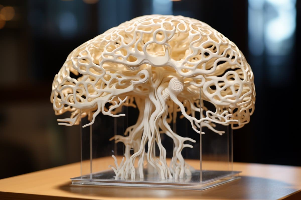
Researchers have achieved a major milestone by creating the world’s first 3D-printed brain tissue that mimics the behavior and growth of natural brain tissue. This advancement represents a significant step forward in the study of neurological and neurodevelopmental disorders. The innovative 3D-printing technique involves using a horizontal layering method and a softer bio-ink, enabling neurons to connect and form networks similar to those found in the human brain. The precise control over cell types and arrangements offers unprecedented opportunities to investigate brain functions and disorders in a controlled environment, providing new avenues for drug testing and understanding brain development and diseases like Alzheimer’s and Parkinson’s. Key Facts: The 3D-printed brain tissue can form networks and communicate through neurotransmitters, similar to human brain interactions. This new printing method allows for precise control over cell types and arrangements, surpassing the capabilities of traditional brain organoids. The technique is accessible to many labs, not requiring special equipment or culture methods, and can significantly impact the study of various neurological conditions and treatments.
Source: University of Wisconsin
The researchers at the University of Wisconsin–Madison have made a groundbreaking discovery by developing the first 3D-printed brain tissue that can grow and function like natural brain tissue. This achievement has noteworthy implications for scientists researching the brain and working on treatments for a wide range of neurological and neurodevelopmental disorders, such as Alzheimer’s and Parkinson’s disease. “This could be a hugely powerful model to help us understand how brain cells and parts of the brain communicate in humans,” remarked Su-Chun Zhang, a professor of neuroscience and neurology at UW–Madison’s Waisman Center.  “Our tissue stays relatively thin and this makes it easy for the neurons to get enough oxygen and enough nutrients from the growth media,” Yan says. Credit: Neuroscience News“It could change the way we look at stem cell biology, neuroscience, and the pathogenesis of many neurological and psychiatric disorders.” Zhang and Yuanwei Yan, a scientist in Zhang’s lab, explained that previous attempts to print brain tissue were hindered by limitations in the printing methods used. The group behind the new 3D-printing process shared their method in the journal Cell Stem Cell. Instead of the traditional approach of stacking layers vertically, the researchers opted for a horizontal orientation. They placed brain cells, neurons developed from induced pluripotent stem cells, in a softer “bio-ink” gel compared to prior attempts. “The tissue still has enough structure to hold together, but it is soft enough to allow the neurons to grow into each other and start communicating,” Zhang highlighted. The cells are arranged next to each other similar to pencils laid out on a tabletop. “Our tissue stays relatively thin and this makes it easy for the neurons to get enough oxygen and enough nutrients from the growth media,” Yan stated. The positive outcomes are evident as the cells are able to establish connections both within each printed layer and across layers, forming networks that resemble those found in human brains. The neurons communicate, send signals, interact with each other through neurotransmitters, and even form proper networks with added support cells in the printed tissue. “We printed the cerebral cortex and the striatum and what we found was quite striking,” Zhang remarked. “Even when we printed different cells belonging to different parts of the brain, they were still able to communicate in a very specific way.” The printing technique offers precision — control over the types and arrangement of cells — not found in brain organoids, miniature organs used to study brains. The organoids grow with less organization and control. “Our lab is very special in that we are able to produce pretty much any type of neurons at any time. Then we can piece them together at almost any time and in whatever way we like,” Zhang explained. “Because we can print the tissue by design, we can have a defined system to look at how our human brain network operates. We can look very specifically at how the nerve cells communicate with each other under certain conditions because we can print exactly what we want.” This specificity provides flexibility. The printed brain tissue could be used to study signaling between cells in Down syndrome, interactions between healthy tissue and neighboring tissue affected by Alzheimer’s, testing new drug candidates, or observing brain growth. “In the past, we have often looked at one thing at a time, which means we often miss some critical components. Our brain operates in networks. We want to print brain tissue this way because cells do not operate by themselves. They communicate with each other. This is how our brain operates, and it has to be studied all together in this manner to truly comprehend it,” Zhang emphasized. “Our brain tissue could be used to study almost every major aspect of what many people at the Waisman Center are working on. It can be used to delve into the molecular mechanisms underlying brain development, human development, developmental disabilities, neurodegenerative disorders, and more.” The new printing technique should also be accessible to many labs. It does not require special bio-printing equipment or complex culture methods to maintain the tissue’s health and can be analyzed in depth with microscopes, standard imaging techniques, and commonly used electrodes in the field. The researchers aim to explore the potential for specialization, further improving their bio-ink and refining their equipment to enable specific orientations of cells within the printed tissue. “Currently, our printer is a commercialized benchtop one,” Yan noted. “We can make specialized enhancements to help us print specific types of brain tissue on-demand.” Funding: This study received support from NIH-NINDS (NS096282, NS076352, NS086604), NICHD (HD106197, HD090256), the National Medical Research Council of Singapore (MOH-000212, MOH-000207), Ministry of Education of Singapore (MOE2018-T2-2-103), Aligning Science Across Parkinson’s (ASAP-000301), the Bleser Family Foundation, and the Busta Foundation. About this neurotech research newsAuthor: Emily Leclerc
Source: University of Wisconsin
Contact: Emily Leclerc – University of Wisconsin
Image: The image is credited to Neuroscience NewsOriginal Research: Open access.
“3D bioprinting of human neural tissues with functional connectivity” by Su-Chun Zhang et al. Cell Stem CellAbstract3D bioprinting of human neural tissues with functional connectivityHighlightsFunctional human neural tissues assembled by 3D bioprintingNeural circuits formed between defined neural subtypesFunctional connections established between cortical-striatal tissuesPrinted tissues for modeling neural network impairmentSummaryProbing how human neural networks operate is hindered by the lack of reliable human neural tissues amenable to the dynamic functional assessment of neural circuits. We developed a 3D bioprinting platform to assemble tissues with defined human neural cell types in a desired dimension using a commercial bioprinter.The printed neuronal progenitors differentiate into neurons and form functional neural circuits within and between tissue layers with specificity within weeks, evidenced by the cortical-to-striatal projection, spontaneous synaptic currents, and synaptic response to neuronal excitation.Printed astrocyte progenitors develop into mature astrocytes with elaborated processes and form functional neuron-astrocyte networks, indicated by calcium flux and glutamate uptake in response to neuronal excitation under physiological and pathological conditions.These designed human neural tissues will likely be useful for understanding the wiring of human neural networks, modeling pathological processes, and serving as platforms for drug testing.
Technology Breakthrough: 3D-Printed Brain Tissue that Works Like Human Brain – Neuroscience News


